
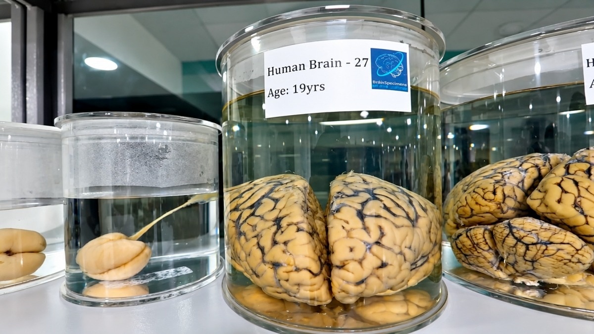
IIT Madras started a project to map 100 human brains in 3D, from foetal stages to old age. Prime Minister Narendra Modi launched this initiative to help researchers understand brain development and disease. The centre will use specialised imaging to create these maps, providing a new resource for scientists worldwide.
What does a human brain look like when you zoom past its anatomy and examine its cellular landscape in three dimensions? IIT Madras is attempting to answer that question by mapping more than 100 human brains, from the earliest stages of foetal development to old age, including brains affected by disease.
The ambitious project, launched by Prime Minister Narendra Modi, aims to create what the Sudha Gopalakrishnan Brain Centre (SGBC) at IIT Madras describes as the world's largest collection of three-dimensional human brain maps at cellular resolution.
The initiative could help researchers understand how the brain develops, ages and changes during disease, creating a publicly accessible resource for scientists and medical professionals worldwide.
The centre will use specialised imaging technology and high-performance computing to transform entire post-mortem human brains into detailed digital 3D maps.
The project will bring together more than 200 researchers, engineers and technicians, over 10 medical institutions across India, and more than 30 leading researchers from over 10 countries.
A NEW WINDOW INTO STROKE-DAMAGED BRAINS
During his visit, PM Modi also released C-STROKE, which IIT Madras describes as the world's first human ischaemic stroke atlas.
The resource connects conventional medical imaging with microscopic examinations of the same stroke-affected brain, helping scientists investigate what happens inside brain tissue when its blood supply is interrupted.
An ischaemic stroke occurs when a blood vessel supplying the brain becomes blocked, depriving brain cells of oxygen and nutrients.
Doctors typically identify stroke-related damage using magnetic resonance imaging (MRI), which captures structures at a millimetre scale. C-STROKE bridges these clinical images with histology, the microscopic examination of tissue, at micron-level resolution.
The atlas contains more than 1,000 annotated brain sections from a stroke-affected brain. It maps neuronal health, inflammation, damage to the blood-brain barrier, oxygen deprivation and injury to nerve fibres.
By linking these cellular changes to patterns visible on MRI scans, researchers could gain a better understanding of the biological processes behind stroke damage and identify potential targets for future treatments.
The atlas was generated using the Param Shakti supercomputer, supported by the Ministry of Electronics and Information Technology. Its open-access viewer and analytical tools will allow researchers worldwide to explore the data.
BUILDING A GLOBAL RESOURCE FOR BRAIN RESEARCH
C-STROKE builds on the centre's earlier work, including DHARANI, released in December 2024, which contains 5,132 sections from five foetal brains.
The new initiative aims to expand such resources across the human lifespan, offering researchers a more comprehensive view of the brain's cellular organisation.
Prof. Mohanasankar Sivaprakasam, head of SGBC, said the project combines brain science, medicine, engineering and computing, with the potential to influence neuroscience and neurological medicine for decades.
By making these detailed maps publicly available, IIT Madras hopes to give researchers a new way to study the human brain, one cell at a time.