
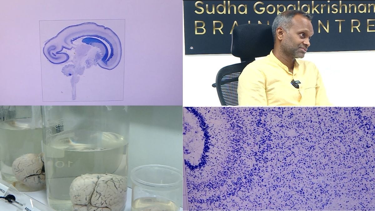
Prime Minister Narendra Modi launched the Sudha Gopalakrishnan Brain Centre at IIT Madras on October 11. Started in a former car park during the 2020 pandemic, the facility now maps human brains at cell resolution. Professor Mohanasankar Sivaprakasam said, "The human brain is one of the most complex organs."
The money arrived in February 2020. By March, the campus gates had closed.
The project had not yet switched on a single machine. The world had gone indoors, and a team of engineers and neuroscientists at IIT Madras was left holding an idea so large that no machine in the world could yet carry it out: to see the human brain cell by cell, from the earliest weeks of life to old age.
They carried on. The laboratory took shape in what had once been a car park.
Professor Mohanasankar Sivaprakasam, who heads the project, says everyone on the team caught Covid-19 along the way, and he jokes that he caught every variant going. Yet, by 2022, the centre was inaugurated, and its machines were running.
On October 11, Prime Minister Narendra Modi launched the Sudha Gopalakrishnan Brain Centre's mission to build 3D maps of more than 100 human brains at cell resolution, IIT Madras announced. This is the story of how it began.
THE ORGAN WE CAN SEE ONLY IN OUTLINE
The brain is the most intricate organ in the body, and the one that science understands least.
"The human brain is one of the most complex organs and one of the least understood," Professor Mohanasankar told India Today Digital in an exclusive interview at the Sudha Gopalakrishnan Brain Centre, IIT Madras. Heart disease, he points out, is far better understood, which is why cardiology has been pushed to extraordinary levels. Neurology and neurosurgery are held back by the gap in our knowledge of the brain itself.
The reason is simple. The brain hides inside the skull. Doctors can study it with an MRI scan, which uses magnetic fields to picture the inside of the body, or by recording its electrical activity. But an MRI can see only down to about a millimetre. A brain cell, called a neuron, measures around 10 microns, and a micron is a thousandth of a millimetre.
By Professor Mohanasankar's reckoning, that is a tenth of the width of a human hair.
The Sudha Gopalakrishnan Brain Centre works at that finer scale. It images whole brains at cell resolution, down to micron-level detail, which is about a thousand times finer than what an MRI can see.
It is a little like watching a city from a passing aeroplane at night. You can tell where the lights are bright, but not who lives in which house. And this city is crowded. The brain holds 80 to 100 billion neurons, along with many more supporting cells, packed into about 1.3 litres. To treat a disease, Professor Mohanasankar says, you need to know which type of cell is affected, and how.
A QUESTION FROM AN ALUMNUS
Every large project begins with a conversation, and this one began with a question from a former student of the institute.
In the middle of the last decade, Kris Gopalakrishnan, Infosys co-founder and an IIT Madras distinguished alumnus, began asking whether the institute could do something big. "The seed and vision for this project started from Kris," says Professor Mohanasankar.
The then director, Professor Bhaskar Ramamurthi, agreed to go ahead. Professor K VijayRaghavan, then the Principal Scientific Adviser to the Government of India, came on board, and in 2018, the decision was taken.
The alumnus, Professor Mohanasankar says, took the first risk by investing in it, and the Principal Scientific Adviser's office backed it too. The centre carries the name of Kris Gopalakrishnan's wife, Sudha, and both attended its inauguration.
It did not start from nothing. The centre grew out of an earlier project to image human brains outside the body at terapixel resolution, which means pictures made of a trillion pixels. By its inauguration, the team had already captured whole-brain, cell-resolution volumes of three developing brains.
A LEAP OVER THE MOUSE
Scientists usually learn on small brains first, and for good reason.
At the time, groups elsewhere were mapping the brains of mice. A mouse brain is smaller than one cubic centimetre. A human brain is about 1,300, which makes it 1,000 to 2,000 times larger, and even a foetal brain is about 100. The team realised that the machinery to map a human brain did not exist anywhere, so they would have to build it.
"We have basically leapfrogged," Professor Mohanasankar says. "Instead of going mouse, monkey, human, we have directly gone to human."
The challenge, he explains, is an engineering one at heart. A single brain must be cut into about 10,000 sections, some as large as 15 centimetres by 20. Each must be photographed.
Then the pieces must be put back together on a computer, which he calls a hard computing problem. The data runs into petabytes, and a petabyte is about a million gigabytes. It must also be fast. At one brain a year, he says, none of them would be around to finish the job.
THE LAB THAT GREW IN A LOCKDOWN
The project was sanctioned in February 2020, and the lockdown arrived in March.
The team had barely begun, Professor Mohanasankar says, with only the administrative formalities done. "This entire technology platform was developed during Covid-19," he says, with new instruments and equipment built from scratch.
Over the next two years, the team built its infrastructure in that car park, turning it into a laboratory. He credits colleagues, including Professor Jayaraj Joseph, for developing many of the technologies. People kept working through the pandemic, and by his account, every one of them fell ill at some point.
The centre was inaugurated on March 19, 2022. Professor V Kamakoti, Director of IIT Madras, said at the time that it would make "deep in-roads in collecting data for brain research."
Professor VijayRaghavan said it would help solve "complex issues that will benefit the world." Its aims included mapping the brain at the level of cells and connections, and training hundreds of students in neuroscience, computing and machine learning. Medical partners included CMC Vellore, NIMHANS and Saveetha Medical College and Hospital.
HOW A BRAIN BECOMES A MAP
The end product is an atlas, a detailed digital map of the brain, much as an atlas of the world maps a country.
The centre acquires brains after death, within eight to 12 hours. That window is called the postmortem interval, the time between death and the preservation of the tissue. Prof. Mohanasankar says the centre has acquired close to 400 brains, from newborns to people in their 90s. Some belonged to people who died of causes unrelated to the brain. Others had brain illnesses such as stroke and neurodegenerative diseases, in which brain cells slowly die, as in dementia.
Light cannot pass through thick tissue, so each brain is sliced into sections only 10 to 20 microns thick. The science of treating and examining such thin slices under a microscope is called histology. Special chemicals then make particular cell types stand out, a technique known as immunohistochemistry. Each slice is photographed, and computers align thousands of images into a single 3D model.
The next layer is molecular. Teams are now working on what scientists call omics, which reads the molecules inside cells. It will be done for selected regions only, because doing it for a whole brain is too costly and slow.
THE MAPS SO FAR, AND THE ROAD AHEAD
The first map the centre released was DHARANI, an atlas of the foetal brain in the second trimester, the middle three months of pregnancy.
It appeared at the turn of 2024 and 2025, and Prof. Mohanasankar stresses that all the data is fully public. In June, the centre released a map of the brainstem, the stalk that joins the brain to the spinal cord and keeps vital functions such as breathing going.
Then came C-STROKE on October 11. IIT Madras describes it as the world's first atlas of a human brain damaged by an ischaemic stroke, which is a stroke caused by a blocked blood vessel. It links MRI scans taken before death to microscope-level sections of the same brain, and holds more than 1,000 annotated sections. They show inflammation, low oxygen, damage to nerve fibres and leakage of the blood-brain barrier, the protective lining of the brain's blood vessels.
The maps were generated on the Param Shakti supercomputer, supported by the Ministry of Electronics and Information Technology, and an open-access viewer lets researchers anywhere study them. Prem Watsa, an IIT Madras distinguished alumnus, supports the research.
In August, Professor Mohanasankar said the centre was about halfway to its goal. He says a minimum of 130 to 140 brains is needed to capture enough variety across ages and diseases. Next come the third trimester and newborn brains, and he hopes to finish within a year and a half to two years. When done, he says, it will be the first cell-resolution atlas of the human brain from foetus to adult in the world.
A RARE KIND OF SUPPORT
Large science projects in India are usually funded by the government, and that limits how far they can go.
Here, Professor Mohanasankar says, private and philanthropic support has carried the centre in a significant way. "That has not happened before in India for a large global science project," he says. The mission now involves more than 200 researchers, engineers and technicians, over 10 medical institutions in India and more than 30 researchers from over 10 countries.
Kris Gopalakrishnan called the Prime Minister's visit "a great inspiration for the scientists working on this mission", and hoped it would encourage more philanthropists to back Indian research.
A map is not a cure, and these atlases are research tools. Yet, Professor Mohanasankar expects them to matter for neuroscience and neuromedicine for decades. The lights went off on a campus in March 2020. Six years later, in a laboratory that grew through a lockdown, they are coming on again, one cell at a time.