
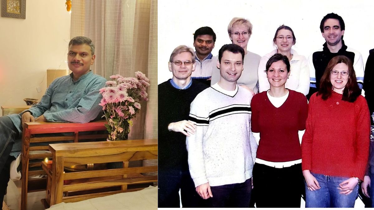
In 2001, Indian researcher Suneel Kateriya identified key light-sensing proteins in an alga while working in Germany. These findings helped Karl Deisseroth, Peter Hegemann and Georg Nagel win the 2026 Medicine Nobel. Kateriya said, "Fundamental science and academic excellence are needed for bigger innovations." His work remains a vital discovery.
In 2001, in Professor Peter Hegemann's lab in Germany, a doctoral student from India searched an alga's gene records and found two light-sensing proteins. Professor Suneel Kateriya, now at Jawaharlal Nehru University (JNU), calls it one contribution to a long journey, and that journey has just won a Nobel Prize.
Somewhere in a freshwater pond, a green speck is swimming towards the Sun. The alga Chlamydomonas is a single cell about 0.015 millimetres across. It has no brain and no nerves, yet it carries an eyespot only about a micrometre wide, which finds light and follows it.
For decades, scientists wanted to know how this speck sees. In 2001, in Regensburg, Germany, a young researcher from India went looking for the answer in the alga's gene records. Hegemann's own 2013 review records the moment plainly. It says that in 2001, Suneel Kateriya, in the Hegemann group, identified new DNA sequences that encoded large microbial-type rhodopsins. The team later named the two genes channelrhodopsin-1 and channelrhodopsin-2.
On October 5, the Nobel Assembly awarded the 2026 Prize in Physiology or Medicine to Karl Deisseroth, Peter Hegemann and Georg Nagel for the discovery of light-gated ion channels and for optogenetics. That is the method that switches chosen brain cells on and off with light. The Nobel Committee's own explainer lists two papers among its key references, and Kateriya is a co-author on both.
He is careful about how his part is told. "Fundamental science and academic excellence are needed for bigger innovations," he told India Today Digital. The discovery, he says, was teamwork led by his supervisor. We also asked three other Indian scientists, from BITS Pilani and IIT Madras, what the prize honours and what still stands in its way.
A QUESTION ABOUT AN ALGA
The question was never about the brain. "We had a fundamental question: how is this alga sensing light, and where are the genes?" Kateriya said. "We never thought it would be used for controlling the human brain."
An early trace of the work is in print. A minireview in the Journal of Phycology, by Hegemann, Markus Fuhrmann and Kateriya, was accepted in August 2001. It reports that new sequences from the alga's genome project code for a seven-segment rhodopsin protein. That sequence, AF385748, is the one the later Science paper analyses. The authors guessed that the protein might act as a light-driven ion channel and noted that the idea still had to be tested.
The test came next. Nagel's experiments showed the proteins work like tiny gates that open in light and change a cell's electrical activity. The first result, on channelrhodopsin-1, appeared in Science in June 2002. The second, on channelrhodopsin-2 (gene AF461397), appeared in PNAS in 2003. Hegemann's lab had spent years failing to purify the alga's light receptors directly. The genes gave them another way in.
A few years later, Deisseroth's team showed that a flash of blue light could switch on nerve cells carrying one of these proteins. Kateriya remembers the training behind it all. "Peter taught me hours and hours, as a PhD student," he said. "That is very important."
HOW A GATE FROM AN ALGA SWITCHES ON A NERVE
A neuron speaks in electricity. It opens tiny gates in its outer wall, and charged particles called ions rush through. Channelrhodopsin is a gate with a difference. The sensor that catches light and the gate that opens are one molecule, so it swings open in light and shuts in the dark.
Put its gene into a neuron, and the neuron begins to obey light. Professor Nishith Gupta, a parasitologist at BITS Pilani's Hyderabad campus, explained why that mattered. "We could stimulate the brain long before optogenetics," he said. "What we lacked was the ability to choose a particular population of neurons and control its activity on the millisecond timescale at which the brain communicates." A millisecond is a thousandth of a second.
Older tools were blunt. Electrodes stimulate a mixture of nearby cells and fibres, and drugs spread wider and last longer. Dr Santhosh Sethuramanujam of IIT Madras's Department of Biotechnology agreed that both lack precision, "indiscriminately affecting multiple cell types across an entire brain region." Light fixes that. "By simply shining light on brain tissue, scientists can switch targeted cells on or off with pinpoint, millisecond precision," he said. And because the gene can be made to work in one cell type alone, only those neurons respond.
THE FEAR THAT LIGHT BROUGHT BACK
In 2012, a team in Susumu Tonegawa's laboratory at MIT used the tool on one of the most private things a brain holds, a memory. Xu Liu, Steve Ramirez and colleagues tagged a small group of hippocampal neurons that had been active while mice learnt to link a particular box with a mild foot shock. Later, in a different, safe box, they switched those cells on with light.
The mice froze, the defensive response tied to the original fright, though no shock was delivered. The controls mattered as much as the result: cells tagged during an experience without a shock did not produce the freezing. "Recording those cells alone could never have established that causal relationship," Gupta said. This was done in mice, and it is not a way to edit human memory.
THE PATIENT AND THE WHITE TABLE
The most striking human result sits on a white table, 80 centimetres square. A notebook, a staple box and a few small tumblers were placed on it, one at a time or in twos and threes, and a patient blinded by retinitis pigmentosa was asked to find them. In this genetic disease, the retina's light-catching photoreceptors slowly die.
In the trial, reported in 2021, doctors injected a virus carrying the gene for a channelrhodopsin called ChrimsonR into the patient's eye. That made the retina's surviving ganglion cells, which carry signals to the brain, sensitive to light. The patient then wore goggles with a camera that noted where the scene changed and projected matching pulses of light onto the retina. Training began about four and a half months after the injection, and about seven months into it the patient began to report improvement.
With the goggles on, the patient perceived, located, counted and touched objects with the treated eye alone. Without them, the patient could detect nothing. It was one patient and a partial recovery, not normal sight. "Blindness is furthest along because the retina is accessible both to gene delivery and to light," Gupta said.
According to its developer, a related retinal therapy, MCO-010, is under regulatory review in the United States and Japan and remains investigational. Its maker, Nanoscope Therapeutics, said on October 1 that Japan had accepted its application for priority review, and it expects decisions in both countries in the first half of 2027. Epilepsy and depression are further off. "Neither condition has an established optogenetic treatment today," Gupta said.
BETWEEN THE LAB AND THE CLINIC
The scientists were as precise about obstacles as they were about promise. Sethuramanujam listed them: "Sensor expression remains transient, efficient expression is limited to specific neuronal populations, and the high light intensities required for activation currently restrict its practical utility in daily life." In plain terms, the protein may not last, cannot yet go into every cell type, and needs light too bright for daily life.
Gupta named a larger barrier: "If I had to name the biggest barrier, it would be achieving safe, durable control of precisely the right cells in the human brain." Light scatters in brain tissue, so deep targets generally need an implanted light source. The gene must also reach the right neurons and stay safe for years. "These problems have to be solved together."
INDIAN LABS, SAME SWITCH
Closer home, three laboratories are using the switch to ask very different questions. At IIT Madras, Sethuramanujam studies how neural circuits encode vision. In earlier postdoctoral work, he used optogenetics to switch on and off neurons that release two chemical signals with opposing effects. It showed that "dual-signal transmission is critical for encoding the direction of object motion within a visual scene", which is how the eye knows which way something moves.
In the same department, Professor Amal Kanti Bera's lab is pointing the tool at glioblastoma, the most aggressive brain cancer. Its cells connect through thin, tube-like bridges called tunnelling nanotubes, which help the tumour survive and resist drugs. The lab is asking what happens to that network when the surrounding neurons are switched on or off. That is a question and a method, not yet a result.
At JNU, Kateriya applies the same tools to ciliopathies, diseases that arise when cilia, the tiny hair-like projections on many of our cells, fail to form or work. He has come full circle, from finding genes in an alga to using the gates they build to study disease.
A single-celled alga evolved a gate to find the light it needs. A student in Regensburg found its genes in a database. A patient in a clinical trial reached for tumblers on a white table. Between those moments lie 25 years of patient, shared science, and the scientists who spoke to us are clear that the journey is not over.